
Ultrasound compared to anatomopathological findings of fetlock
Examination of 37 forelimbs approved that findings of ultrasound changes in shape, continuity and echogenicity of tendons, ligaments, joint capsule, articular cartilage and bony surfaces of the metacarpophalangeal joint were related to the macroscopic and histologic findings.
At a slaughter house, 37 forelimbs were selected which obtained physical changes at inspection and palpation. No medical history of these horses was taken. The control group was the contralateral forelimb.
Changes of the MCP joint structures visualized with ultrasound were documented. Next, forelimbs were dissected and exposed to macroscopic study, to observe changes in size, shape, consistency, color and presence of adhesions. Then soft tissue samples were used for histopathology.
54 abnormal structures were identified on ultrasonographic images and confirmed by anatomopathological investigation. Ultrasound examination appeared to be beneficial for detecting pathology of 3 different categories:
- Joint Capsule and Articular Cartilage
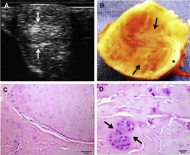
Osteoarticulair changes on ultrasonographic images, included: loss of the anechoic space and irregularity of subchondral bone surface. This was linked to a strong occurrence of linear grooves in the metacarpal condyle and histologic changes: fibrillation and eburnation of the articular cartilage. - Ligaments and Tendons
Hyperechogenic areas in ultrasound images were mainly typified by white fibrous tissue and histologically by multifocal severe cartilaginous metaplasia. See image below. - Proximal Sesamoid Bones
> G. De Bastiani et al. / Journal of Equine Veterinary Science 34 (2014) 1218–1225. All rights reserved to 2014 Elsevier Inc. Click here for the J-EVS summary Click here for the full-text