

Ossification in the shoulder region and hopping-type lamenes
In this case study describes Dr. Sue Dyson an event horse which had developed a hopping-type forelimb lameness. On a firm surface lungeing on the left circle the horse shortened its left forelimb stride length, was unbalanced and marginally lame. This hopping-type left forelimb lameness was apparent when ridden and was not influenced by any local analgesic technique. Because of multilimb lameness the horse was euthanized and post mortem examination was performed.
Radiological assessment pre-euthanasia showed a thin, broad, mineralized plate in the left shoulder between the biceps brachii and brachiocephalicus muscles. This hyperechoic mass cranial to the intertubercular bursa was also seen in ultrasound assessment. There was no involvement of the intertubercular bursa or biceps tendon or muscles.
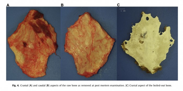
Post-mortem examination revealed a rhomboid shaped and convex plate of bone approximately 3 to 4 mm thick, 11 cm long by 10 cm wide (see image below). It was easily dissected and no abnormalities of the muscles, tendon and bursa were detected. This phenomenon is called heterotopic ossification and also known as myositis ossificans. It is a commonly described condition in humans, typified by the development of bone at sites where bone does not normally exist. The cause is unknown.
Dr. Sue Dyson highlights the value of evaluating lame horses under different conditions, because the lameness can vary depending on the circumstances. Lameness seen in hand or on the lunge may not be due to the same cause as lameness seen when ridden
> From: Dyson, J. Equine. Vet. Sci. 34 (2014) 532-537. All rights reserved to Elsevier Inc.. Click here for the online summary.